Our Health Library information does not replace the advice of a doctor. Please be advised that this information is made available to assist our patients to learn more about their health. Our providers may not see and/or treat all topics found herein.
Ear Examination
Test Overview
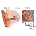
An ear exam is a thorough check of the ears. It is done to screen for ear problems, such as hearing loss, ear pain, discharge, lumps, or objects in the ear. An ear exam can find problems in the ear canal, eardrum, and middle ear. These problems may include infection, too much earwax, or an object like a bean or a bead.
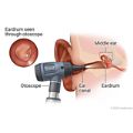
During an ear exam, a tool called an otoscope is used to look at the outer ear canal and eardrum. An otoscope is a handheld tool with a light and a magnifying lens. It also has a funnel-shaped viewing piece with a narrow, pointed end called a speculum. A pneumatic otoscope has a rubber bulb that your doctor can squeeze to give a puff of air into the ear canal. The air helps the doctor to see how the eardrum moves.
Why It Is Done
An ear exam may be done:
- As part of a routine physical exam.
- As part of the process to screen newborns for hearing loss.
- To find the cause of symptoms such as earache, a feeling of pressure or fullness in the ear, or hearing loss.
- To check for excess wax buildup or an object in the ear canal.
- To find the location of an ear infection. The infection may just be in the external ear canal (otitis externa). Or it might be in the middle ear behind the eardrum (otitis media).
- To see how the treatment for an ear problem is working.
How To Prepare
It is important to sit very still during an ear exam. A young child should be lying down with their head turned to the side. Or the child may sit on an adult's lap with the child's head resting securely on the adult's chest. Older children and adults can sit with the head tilted slightly toward the opposite shoulder.
Your doctor may need to remove earwax in order to see the eardrum.
How It Is Done
An ear exam can be done in a doctor's office, a school, or the workplace.
For an ear exam, the doctor uses a special tool called an otoscope to look into the ear canal and see the eardrum.
Your doctor will gently pull the ear back and slightly up to straighten the ear canal. For a baby under 12 months, the ear will be pulled downward and out to straighten the ear canal. The doctor will then insert the pointed end (speculum) of the otoscope into the ear and gently move the speculum through the middle of the ear canal to avoid irritating the canal lining. The doctor will look at each eardrum (tympanic membrane).
Using a pneumatic otoscope lets your doctor see what the eardrum looks like. It also shows how well the eardrum moves when the pressure inside the ear canal changes. It helps the doctor see if there is a problem with the eustachian tube or fluid behind the eardrum (otitis media with effusion). A normal eardrum will flex inward and outward in response to the changes in pressure.
How It Feels
The physical exam of the ear using an otoscope usually isn't painful. If you have an ear infection, putting the otoscope into the ear canal may cause mild pain.
Risks
The pointed end of the otoscope can irritate the lining of the ear canal. This can often be avoided by putting the otoscope in slowly and carefully. If the otoscope does scrape the lining of the ear canal, it could cause bleeding or infection, but this is rare.
Results
Normal: |
|
|---|---|
Abnormal: |
|
Related Information
Credits
Current as of: October 27, 2024
Author: Ignite Healthwise, LLC Staff
Clinical Review Board
All Ignite Healthwise, LLC education is reviewed by a team that includes physicians, nurses, advanced practitioners, registered dieticians, and other healthcare professionals.
Current as of: October 27, 2024
Author: Ignite Healthwise, LLC Staff
Clinical Review Board
All Ignite Healthwise, LLC education is reviewed by a team that includes physicians, nurses, advanced practitioners, registered dieticians, and other healthcare professionals.
This information does not replace the advice of a doctor. Ignite Healthwise, LLC disclaims any warranty or liability for your use of this information. Your use of this information means that you agree to the Terms of Use and Privacy Policy. Learn how we develop our content.
To learn more about Ignite Healthwise, LLC, visit webmdignite.com.
© 2024-2025 Ignite Healthwise, LLC.


